
Dental Cleaning & Prevention
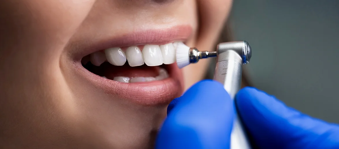
A preventive program is when you, your dentist, and the dental team work together to keep your natural teeth and supporting structures healthy. This means stopping dental issues from happening or getting worse.
The first step in preventing dental problems is taking care of your teeth at home through good oral hygiene and a balanced diet. Then, your dentist and dental hygienist do their part in the office to make sure your oral health stays great.
Prevention also means getting regular dental check-ups, cleanings, and x-rays. Things like sealants and fluoride treatments also help prevent problems by keeping your teeth safe.
By focusing on prevention, you can avoid big and expensive dental problems and keep your smile healthy, confident, and beautiful.
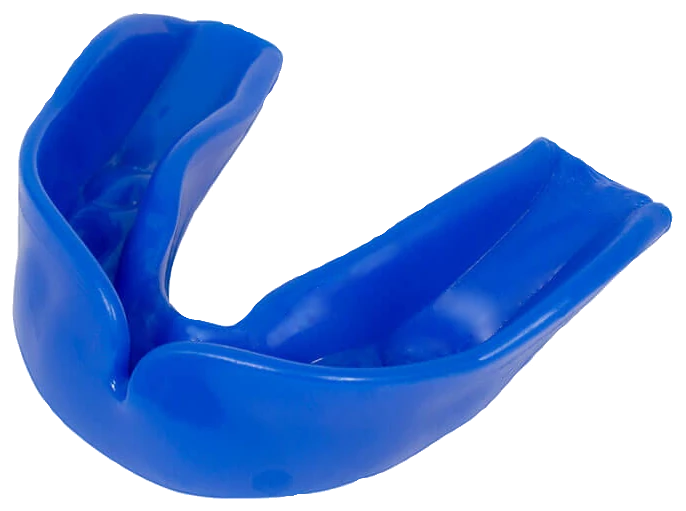 Caring for Athletic Mouth Guards
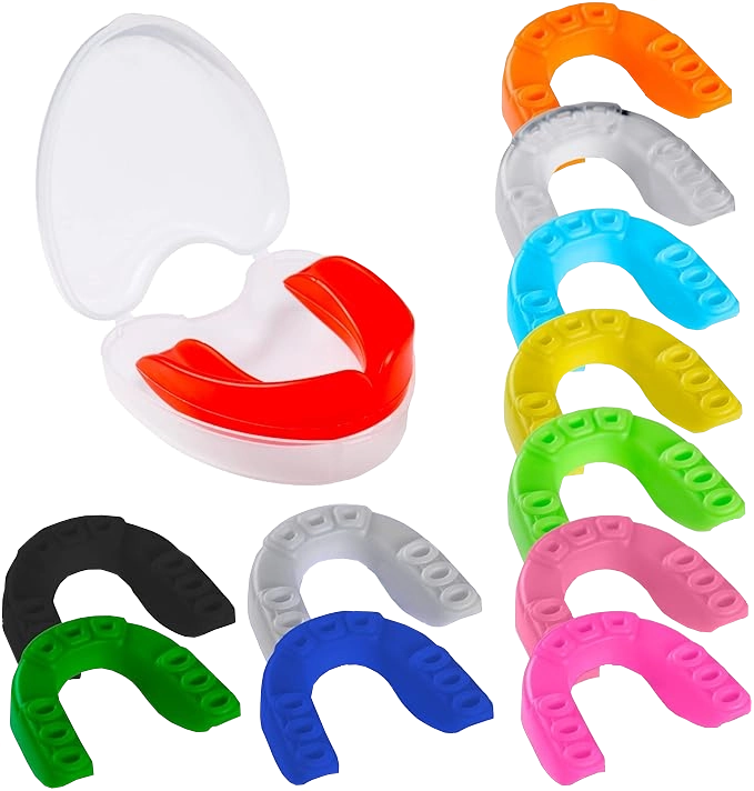
TAKING CARE OF SPORTS MOUTH GUARDS
Participating in sports can put your teeth and orthodontic devices at risk, especially for young people. If you or a family member is into sports, it's crucial to shield yourself from potential harm with a special mouth guard made for sports. People often forget how important mouth guards are for sports, but using them can make you up to sixty times less likely to get oral injuries compared to those who don't use them.
When you use, store, and take care of sports mouth guards correctly, they provide solid protection for your smile.
Picking a good mouth guard is key — it should be easy to clean, resistant to tearing, comfy, and not get in the way of speaking or breathing.
Here are some tips for looking after your mouth guard:
Cleaning— Before and after each use, clean the mouth guard with a toothbrush and toothpaste. This gets rid of bacteria and makes it feel clean and fresh.
Rinsing — Every so often, give the mouth guard a good clean and rinse with soap and lukewarm water to get rid of any debris thoroughly.
Storage — When you're not using it, the best way to keep your mouth guard safe is to keep it in a sturdy case. This protects it from getting crushed or broken.
Replacement — Over time, mouth guards wear out and can't protect your teeth as well. Pay attention to its condition and ask your dentist when it's time for a new one.
If you've got any questions or worries about sports mouth guards, don't hesitate to get in touch with your dentist.
 Simple Tooth
Extractions
SIMPLE TOOTH REMOVAL
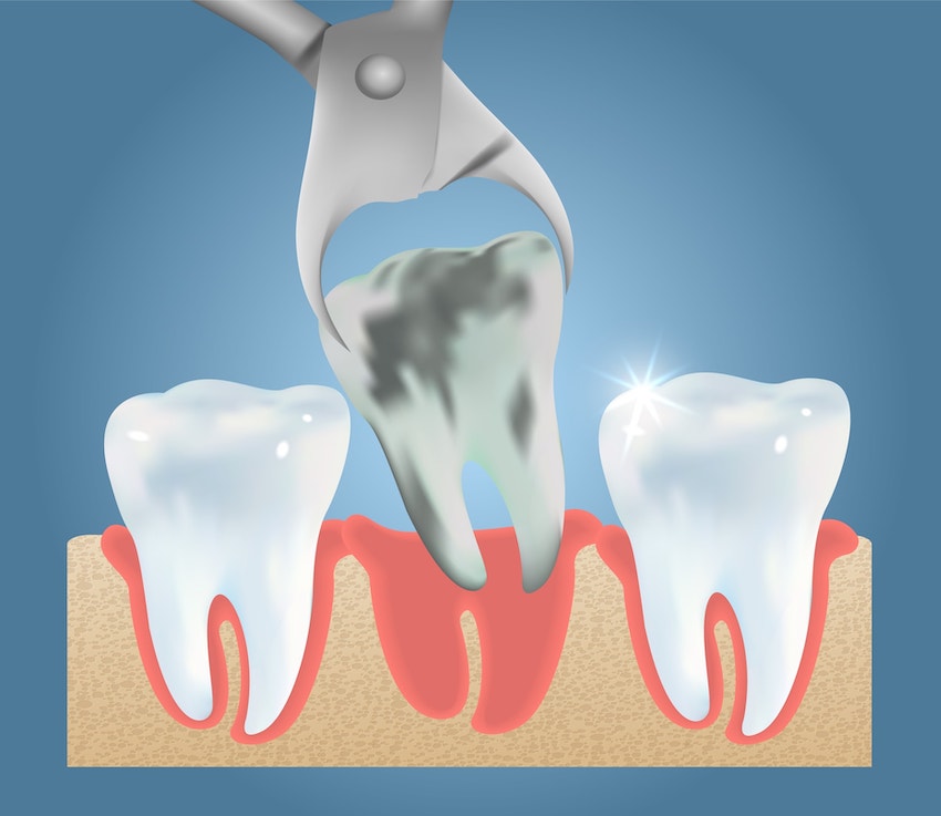
Sometimes, if you're dealing with really sensitive teeth or have a serious gum disease, you might need to have a tooth taken out. But don't worry – with a simple tooth extraction, you won't need a big surgery. The dentist can get rid of the problem tooth safely.
Why You Might Need a Tooth Removed
There are a bunch of situations where a simple extraction can help ease pain or get you ready for other dental work. Here are some common reasons:
- When gum disease has made your tooth roots loose.
- If you've got extra teeth or baby teeth getting in the way of your adult teeth.
- When you need to get ready for braces.
- If a tooth is broken or not formed properly.
- When tooth decay is really bad and can't be fixed with a root canal.
How a Tooth Gets Removed
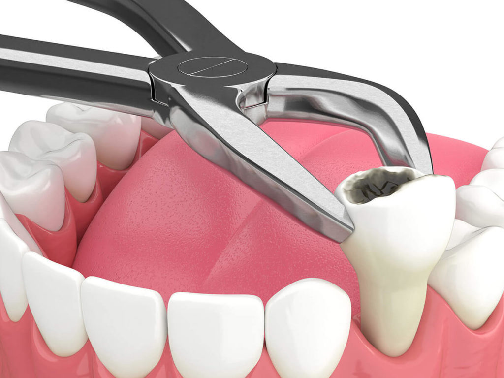
First, the dentist will take X-rays of the tooth (or teeth) to plan how to do the extraction. To make sure you don't feel pain, they'll give you something to numb the area. Then, using a tool called an elevator, they'll gently lift the tooth and loosen the gum tissue around it. After that, they'll use forceps to wiggle the tooth until it comes out of the gums. Sometimes, a tough tooth might not want to come out easily. In these cases, the dentist might have to break the tooth into smaller pieces to remove it.
Once the tooth is out, the dentist will put some gauze in the hole and have you bite down on it. If needed, they might put stitches to close the hole.
If you're sick a week before your extraction or on the day of, let our office know. We might need to change your appointment. If you have any questions or worries, just get in touch with us.
 X-Rays
X-RAYS: CLEARER INSIGHT INTO DENTAL HEALTH
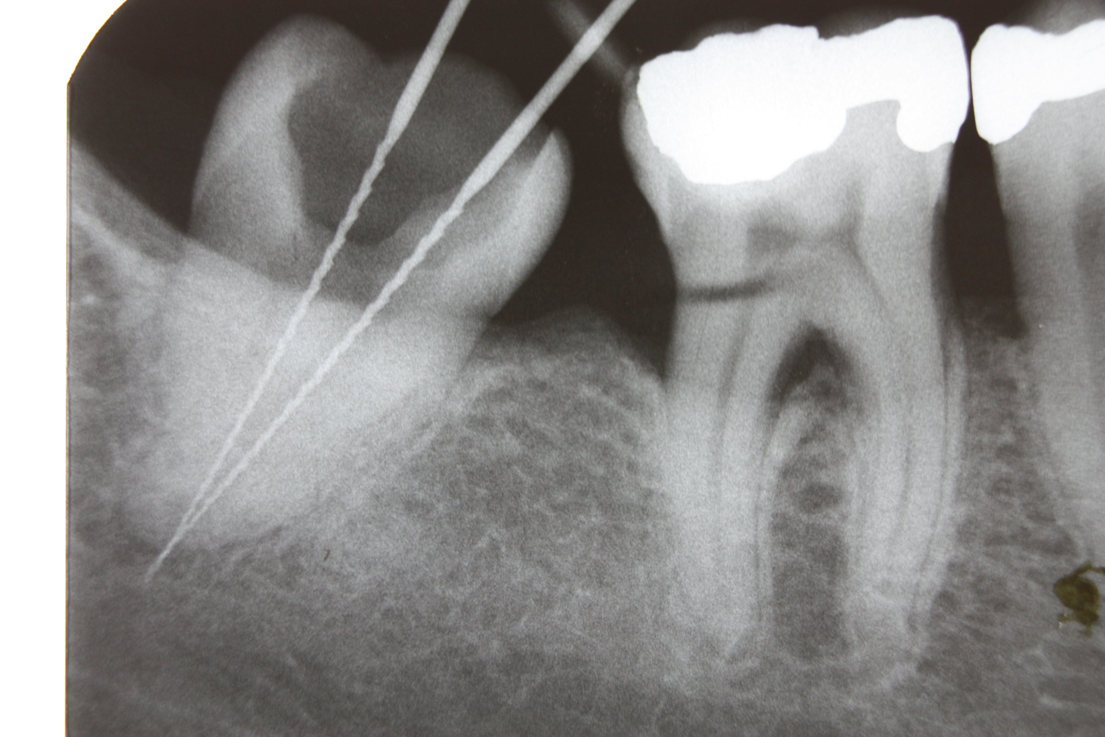
Digital radiography, also known as digital X-ray, is the cutting-edge technology utilized for dental X-rays. This method employs an electronic sensor instead of traditional X-ray film to capture and store images on a computer. The digital image can be instantly examined and enlarged, aiding dentists and dental hygienists in spotting issues more effectively. Moreover, digital X-rays lower radiation exposure by 80-90% compared to conventional dental X-rays, which already have low radiation levels.
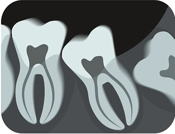
Dental X-rays play a crucial role as preventive and diagnostic tools, providing essential insights not discernible during regular dental exams. Dentists and dental hygienists rely on these X-rays to identify hidden dental irregularities accurately and develop precise treatment plans. Without X-rays, potential problem areas might remain unnoticed.
Dental X-rays can unveil a range of conditions:
- Abscesses or cysts.
- Bone loss.
- Cancerous and non-cancerous tumors.
- Decay between teeth.
- Developmental abnormalities.
- Poor tooth and root positions.
- Problems beneath the gum line or within a tooth.
Detecting and addressing dental issues in their early stages can save you time, money, unnecessary discomfort, and preserve your teeth!
Are dental X-rays safe?
Natural radiation is present in our environment, and digital X-rays emit considerably lower radiation levels compared to traditional ones. Not only are digital X-rays better for patient health and safety, but they are also quicker and more comfortable, reducing your time at the dental office. Additionally, digital images don't require development, eliminating the need to dispose of harmful waste and chemicals into the environment.
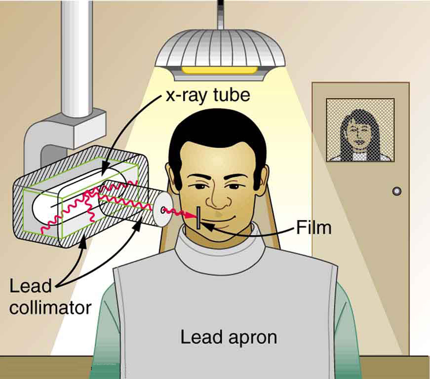
Despite the low radiation levels of digital X-rays, dentists take precautions to minimize patient exposure. This includes only taking necessary X-rays and using lead apron shields for protection.
How often should dental X-rays be taken?
The necessity for dental X-rays varies based on each individual's dental health needs. Recommendations for X-rays depend on factors such as medical and dental history, symptoms, age, and disease risk. New patients often undergo a full mouth series of dental X-rays, which remains useful for three to five years.
For new patients, a full mouth series of X-rays is recommended. Typically, this series remains useful for three to five years. Bite-wing X-rays (images of upper and lower teeth biting together) are taken during check-up visits and suggested once or twice a year to detect emerging dental issues.
 Fluoride Treatment
FLUORIDE TREATMENT: GUARDING AGAINST TOOTH DECAY
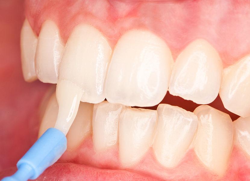
Fluoride stands as the most effective agent in the battle against tooth decay. This mineral is naturally present in various amounts in almost all foods and water supplies. The benefits of fluoride have been recognized for over half a century and are endorsed by numerous health and professional organizations.
Fluoride operates in two ways:
- Topical Fluoride: This strengthens teeth once they've grown by seeping into the outer layer of tooth enamel, enhancing their resistance to decay. We encounter topical fluoride through dental products containing fluoride like toothpaste, mouth rinses, and gels. Dentists and dental hygienists often suggest professional fluoride application twice a year during dental check-ups, especially for children.
- Systemic Fluoride: This fortifies both erupted teeth and those still developing under the gums. We receive systemic fluoride from various foods and our community water sources. It's also available as supplements in drop or gel form, prescribed by dentists or physicians. Generally, fluoride drops suit infants, while tablets are more fitting for children and teens. Monitoring fluoride intake is crucial, as excessive consumption during tooth development could lead to fluorosis (white spots on teeth).
Though many get fluoride from food and water, sometimes it's insufficient to thwart decay. Your dentist or dental hygienist might advise home and/or professional fluoride treatments for the following reasons:
- Deep pits and fissures on tooth chewing surfaces.
- Exposed and sensitive tooth root surfaces.
- Fair to poor oral hygiene habits.
- Frequent consumption of sugar and carbs.
- Inadequate exposure to fluoride.
- Reduced saliva flow due to health conditions, treatments, or medications.
- Recent history of dental decay.
Remember, fluoride alone can't prevent tooth decay! It's vital to brush at least twice daily, floss consistently, have balanced meals, cut down on sugary snacks, and maintain regular dental check-ups.
 i-CAT® 3D Imaging
I-CAT® 3D IMAGING: A NEW DIMENSION IN DENTAL INSIGHT
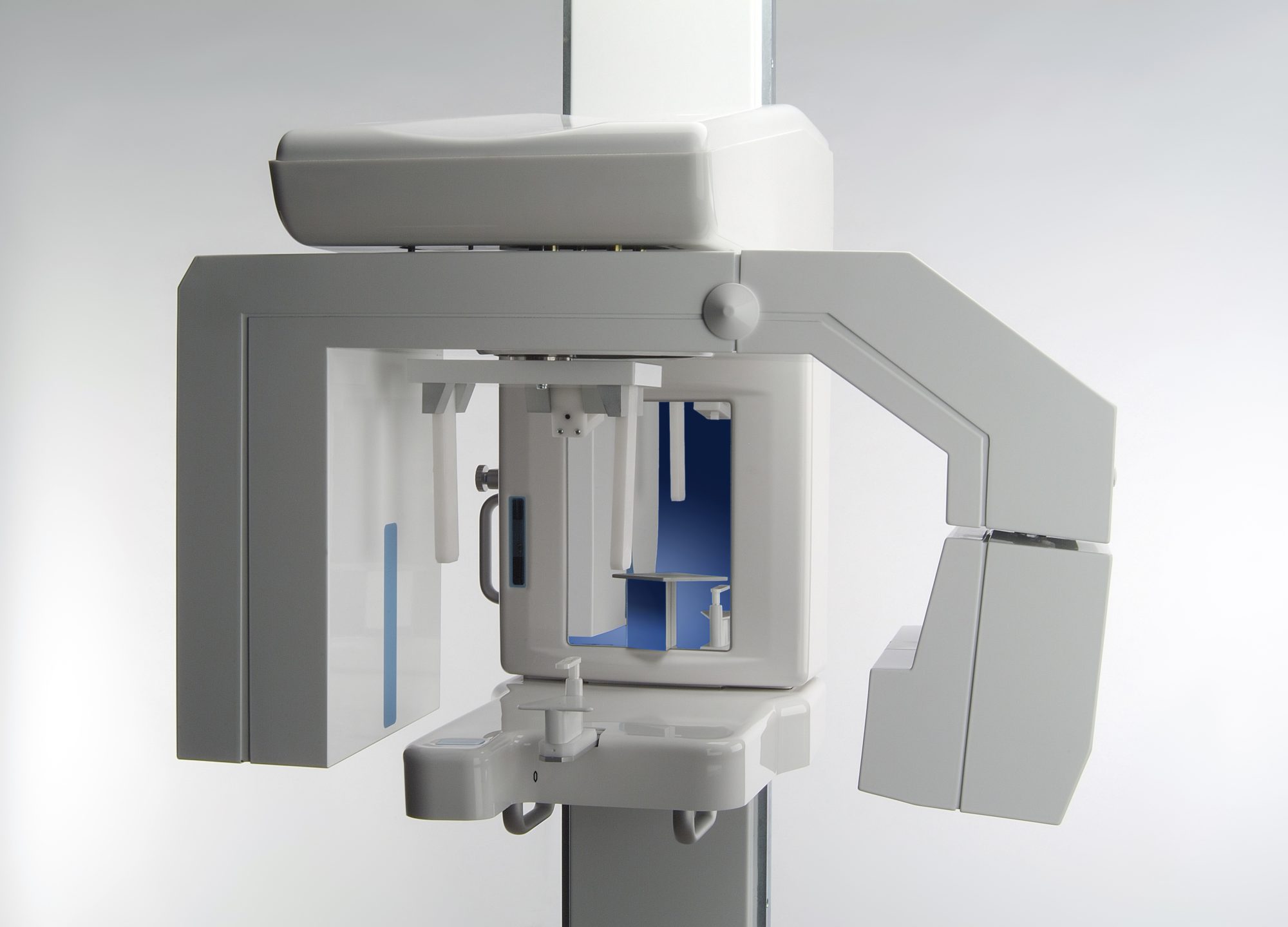
For years, doctors have relied on computerized axial tomography scans (CAT scans) to peer into the human body. These scans, which use X-rays and computers to create cross-sectional and 3D views of internal structures, have become vital tools in medical diagnostics. Think of a knee replacement surgery – it wouldn't proceed without thorough 3D imaging.
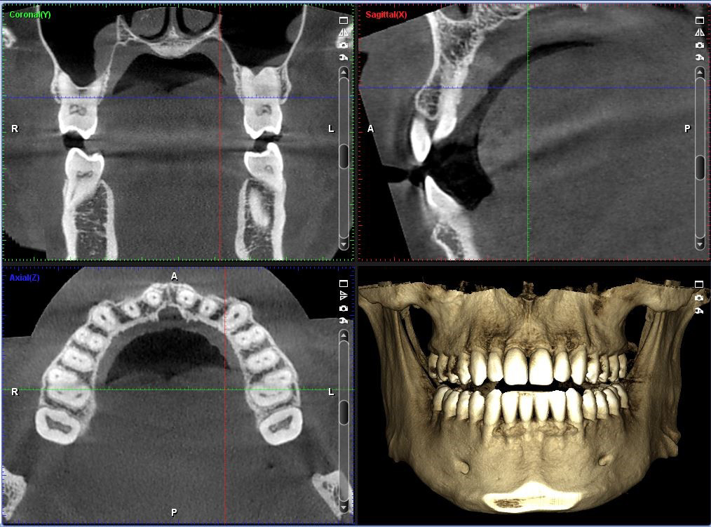
Recently, dentists have embraced 3D imaging techniques and i-CAT® scans to gain a detailed look at the mouth and skull. The advantage of 3D imaging over regular dental X-rays lies in its clarity in depicting bone structure, density, tissues, and nerves.
i-CAT® scans are incredibly efficient, taking less than half a minute to complete. This means they expose the body to far less radiation than traditional bitewing X-rays. Their primary role is aiding in planning dental implant treatment and other oral surgeries.
Dental implants are advanced tooth replacements, but their placement used to be time-consuming. i-CAT® scans have revolutionized this process, significantly reducing implant placement time. It's believed that in the near future, implants could be placed in a single visit thanks to this unique imaging technique.
How are i-CAT® scans utilized?
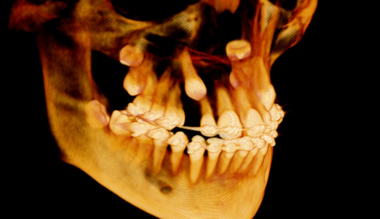
i-CAT® scans offer the benefit of magnifying specific facial areas. Dentists can view cross-sectional "slices" of the jaw, simplifying and expediting treatment planning.
Here are the primary ways i-CAT® scans are used in dentistry:
- Evaluating jawbone quality for implant placement.
- Locating nerve positions.
- Early-stage tumor and disease diagnosis.
- Assessing jawbone density for implant placement.
- Determining optimal implant placement, including angle.
- Pre-planning the entire surgical procedure.
- Selecting precise implant size and type.
- Visualizing tooth orientation and position.
- Observing impacted teeth.
How are i-CAT® scans performed?
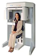
i-CAT® scans are swift and straightforward. At the core of the i-CAT® scanner is a Cone Beam Imaging System. Patients remain seated during the scan. Cone beams take numerous facial pictures, forming an accurate 3D image of the face and jaw's inner workings. The dentist can zoom in on specific regions and examine them from different angles.
Previous patients find the i-CAT® scanner comfortable since they remain seated. The open setup eliminates claustrophobia concerns. The i-CAT® scan is a remarkable tool that minimizes dental treatment costs, reduces treatment time, and enhances surgical outcomes.
For any inquiries about i-CAT® scans or 3D imaging, please reach out to our office.
 Sealants
DENTAL SEALANTS
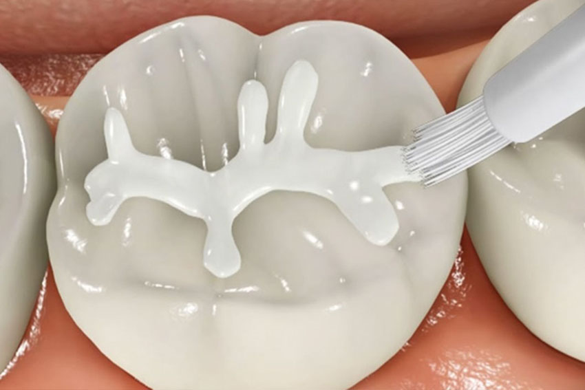
Sealants are thin, plastic coatings that dentists apply to the chewing surfaces of molars, premolars, and deep grooves (known as pits and fissures) on teeth. These deep grooves are where over 75% of dental decay originates. Such teeth are hard to clean and highly prone to decay. Sealants shield teeth by sealing these grooves, creating a smooth surface that's easier to clean.
While sealants can safeguard teeth from decay for many years, it's important to have them regularly checked for wear and chipping during dental visits.
Why are sealants used?
Sealants are recommended for:
- Children and teenagers: As soon as the first permanent back teeth (six-year molars) emerge or at any time between ages 6 and 16 when cavities are common.
- Adults: On tooth surfaces without decay but with deep grooves or depressions.
- Baby teeth: Sometimes, if the teeth have deep grooves or depressions and the child is prone to cavities.
How are sealants applied?
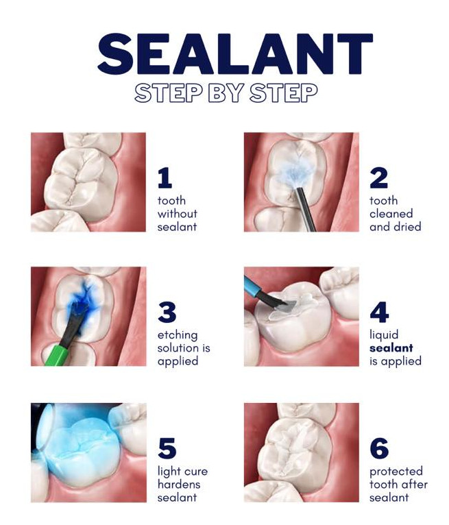
Dentists or dental hygienists can easily apply sealants, and the process typically takes only a couple of minutes per tooth.
The procedure involves:
- Thoroughly cleaning the teeth to be sealed.
- Using cotton to isolate and keep the area dry.
- Applying a special solution to the enamel surface to promote bonding.
- Rinsing and drying the teeth.
- Carefully painting sealant material onto the enamel to cover the deep grooves or depressions.
- Depending on the sealant type, the material will either harden on its own or with a special curing light.
Maintaining proper oral hygiene, a balanced diet, and regular dental check-ups will help prolong the effectiveness of your new sealants.
 Home Care
TAKING CARE OF YOUR SMILE AT HOME
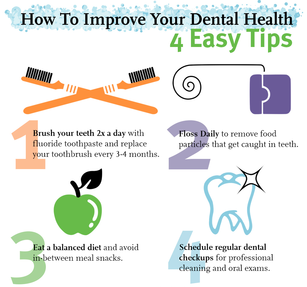
Our main goal is to provide you with a healthy, beautiful smile that lasts a lifetime. Your role in achieving this goal is crucial through your home care routine. Proper home care involves eating balanced meals, limiting snacks, and using the right dental tools to control plaque and bacteria, which lead to dental problems.
Tooth brushing — Brush your teeth at least twice a day, especially before bedtime, with a soft-bristle brush and ADA-approved toothpaste.
- Hold the brush at a 45-degree angle to your gums and gently brush using small, circular motions, making sure the bristles touch your gums.
- Brush the outer, inner, and biting surfaces of every tooth.
- Clean the inside of your front teeth using the brush's tip.
- Don't forget to brush your tongue to remove bacteria and freshen your breath.
- Electric toothbrushes are also recommended. They're easy to use and can efficiently remove plaque. Simply place the brush on your gums and teeth and let it do the work, focusing on a few teeth at a time.
Flossing — Daily flossing is essential for cleaning between teeth and under the gumline. Flossing not only cleans these areas, but also prevents plaque buildup, which can harm gums, teeth, and bone.
- Take about 12-16 inches of dental floss and wrap it around your middle fingers, leaving around 2 inches between them.
- Use your thumbs and forefingers to guide the floss, gently inserting it between teeth with a sawing motion.
- Curve the floss into a "C" shape around each tooth and beneath the gumline. Gently move the floss up and down, cleaning the sides of each tooth.
- Floss holders can be helpful if you find traditional floss difficult to use.
Rinsing — After brushing or meals, it's important to rinse your mouth with water if you can't brush. If you're using an over-the-counter rinse, consult your dentist or dental hygienist to ensure it's suitable for you.
Use other dental aids as advised by your dentist or dental hygienist: interdental brushes, rubber tip simulators, tongue cleaners, irrigation devices, fluoride, medicated rinses, and more can all contribute to effective dental home care.
